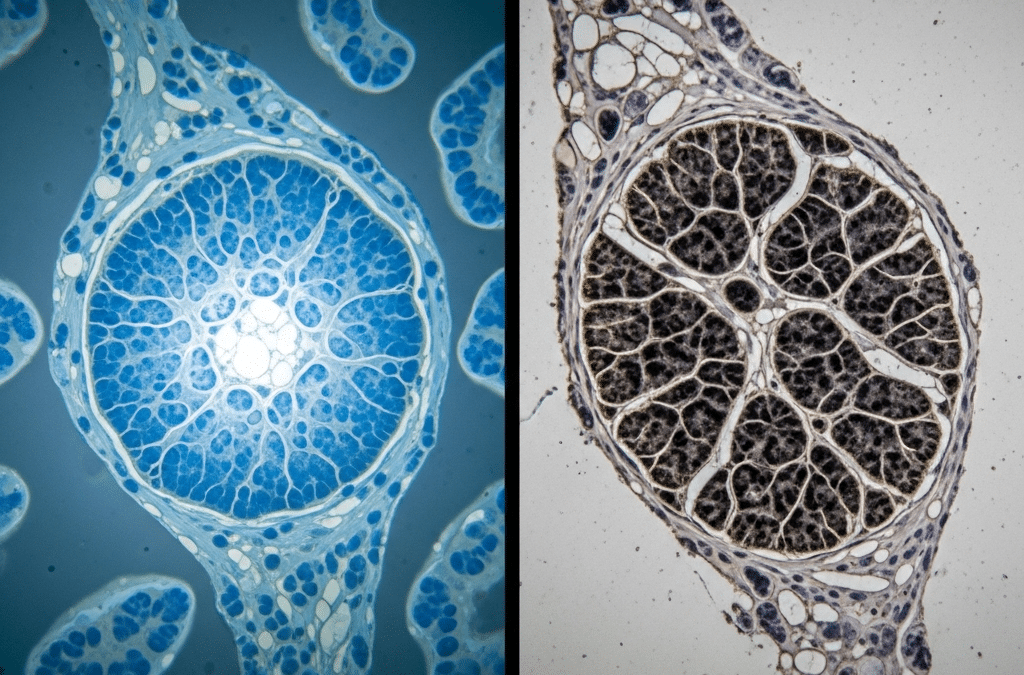
Cryopreserved tissue (often referred to as “fresh frozen” or “flash-frozen”) and standard pathology (Formalin-Fixed Paraffin-Embedded, or FFPE) tissue represent two fundamentally different approaches to tissue preservation, each with distinct advantages and disadvantages, especially concerning downstream applications.
Here’s a breakdown of their differences:
1. Preservation Method:
- Cryopreserved Tissue:
- Method: Rapidly cooled to extremely low temperatures, typically -80°C (in an ultra-low freezer) or -196°C (in liquid nitrogen).1 This process, known as “flash freezing” or “snap freezing,” essentially halts all biological activity (metabolism, degradation) and keeps the tissue in a “native” or “as-is” state.
- Mechanism: Prevents ice crystal formation by rapid freezing, and/or uses cryoprotective agents (CPAs) to minimize cellular damage from ice. The goal is to preserve cellular structures and biomolecules (DNA, RNA, proteins) in their original conformation and activity.2
- Standard Pathology (FFPE) Tissue:
- Method: Involves two main steps:
- Fixation: The tissue is immersed in a chemical fixative, most commonly formalin (a solution of formaldehyde).3 Formalin cross-links proteins, effectively “fixing” the tissue’s structure and preventing degradation by enzymes.4
- Embedding: After fixation, the tissue is dehydrated through alcohol solutions and then infiltrated with and embedded in paraffin wax, creating a solid block.5
- Mechanism: Formalin fixation preserves the tissue’s morphology (shape and structure) exceptionally well, making it ideal for routine microscopic examination by pathologists.6 The paraffin provides a stable matrix for long-term storage at room temperature.
- Method: Involves two main steps:
2. Impact on Biomolecules:
- Cryopreserved Tissue:
- DNA/RNA: Considered the “gold standard” for high-quality DNA and RNA extraction. Nucleic acids remain largely intact and undegraded, making them ideal for sensitive molecular analyses like next-generation sequencing (NGS), PCR, and gene expression profiling (e.g., RNA-Seq).
- Proteins: Proteins largely retain their native conformation, enzymatic activity, and antigenicity (their ability to bind to antibodies). This is crucial for proteomics studies, enzyme assays, and certain immunohistochemistry (IHC) applications where native protein structure is critical.
- Lipids: Lipids are generally well-preserved.
- Standard Pathology (FFPE) Tissue:
- DNA/RNA: Formalin fixation causes cross-linking and fragmentation of nucleic acids. While DNA and RNA can still be extracted, they are often degraded and chemically modified.7 This can make certain molecular analyses more challenging, require specialized kits, and may lead to lower yields or introduce artifacts in sequencing data.
- Proteins: Formalin cross-links proteins, which can alter their native conformation and lead to a loss of enzymatic activity.8 While many antigens can still be detected via IHC after “antigen retrieval” techniques, some are irrevocably masked or denatured, limiting their use for certain protein studies (e.g., studies of post-translational modifications, or some antibodies may not bind effectively).
- Lipids: Lipids are often dissolved or altered during the dehydration and paraffin embedding process.
3. Applications and Strengths:
- Cryopreserved Tissue:
- Molecular Biology: Preferred for almost all cutting-edge molecular analyses, including genomics (whole-genome sequencing, exome sequencing), transcriptomics (RNA sequencing), proteomics (mass spectrometry-based analysis), and epigenomics.
- Personalized Medicine: Essential for personalized drug sensitivity testing (e.g., growing organoids from live tumor cells), developing patient-specific immunotherapies (e.g., T-cell therapies, vaccines), and understanding tumor evolution.
- Viability Studies: Can sometimes retain cell viability (if processed appropriately), allowing for cell culture or xenograft models.
- “Omics” Research: Ideal for comprehensive “omics” studies where high-quality, intact biomolecules are paramount.
- Standard Pathology (FFPE) Tissue:
- Routine Pathology Diagnosis: The gold standard for routine histopathological diagnosis. Pathologists rely on the excellent morphological preservation to identify cell types, tissue architecture, and disease features under the microscope.9
- Immunohistochemistry (IHC): Widely used for IHC, a technique that uses antibodies to detect specific proteins in tissue sections, aiding in diagnosis, prognosis, and treatment prediction.
- Long-Term Storage: FFPE blocks are extremely stable and can be stored at room temperature for decades, making them widely accessible and cost-effective for large historical archives and retrospective studies.10
- Ease of Handling/Transport: Easier to handle and transport compared to frozen tissue, which requires continuous ultra-cold storage.
4. Limitations:
- Cryopreserved Tissue:
- Morphological Artifacts: Ice crystal formation during freezing can sometimes introduce morphological artifacts, making detailed histological examination more challenging compared to FFPE.11
- Logistics: Requires specialized equipment (liquid nitrogen tanks, -80°C freezers) and careful handling to maintain ultra-low temperatures, making collection, storage, and transport more complex and expensive.
- Availability: Less commonly collected and stored than FFPE in routine clinical practice, making large historical cohorts harder to access.
- Standard Pathology (FFPE) Tissue:
- Molecular Degradation: The major limitation is the damage and degradation of DNA, RNA, and proteins, which can complicate or limit advanced molecular analyses.12
- Bias/Artifacts: Fixation can introduce chemical modifications and artifacts that might affect the accuracy of certain molecular assays.13
- Limited for “Live” Assays: Cannot be used for applications requiring viable cells (e.g., cell culture, functional assays).
In Summary:
| Feature | Cryopreserved (Fresh Frozen) Tissue | Standard Pathology (FFPE) Tissue |
| Preservation Method | Rapid freezing (liquid nitrogen, -80°C freezer); halts metabolism. | Formalin fixation (cross-links proteins) followed by paraffin embedding; preserves structure. |
| Biomolecule Quality | High: Intact, native DNA, RNA, and proteins. Gold standard for molecular analysis. | Lower: Degraded, fragmented, and modified DNA/RNA/proteins due to cross-linking. |
| Morphology | Good, but can have freezing artifacts; less ideal for routine microscopic diagnosis. | Excellent: Preserves cellular and tissue architecture for clear microscopic diagnosis. |
| Storage | Requires ultra-cold freezers (-80°C) or liquid nitrogen (-196°C); high maintenance, vulnerable to power outages. | Room temperature; highly stable for decades; easy to store and transport. |
| Primary Use | Advanced molecular profiling (genomics, transcriptomics, proteomics), personalized medicine research, drug testing, biobanking for future technologies. | Routine histopathological diagnosis, immunohistochemistry (IHC), large retrospective studies, general biobanking. |
| Logistics | More complex, expensive, and requires specialized equipment and handling. | Simpler, less expensive, and widely established in clinical labs. |
For cutting-edge research, especially in personalized oncology where detailed molecular insights are crucial, cryopreserved tissue is increasingly preferred as the “gold standard.” However, FFPE remains indispensable for routine diagnosis and large-scale historical studies due to its robust morphological preservation and ease of storage. Ideally, for comprehensive patient care and research, both types of preservation are utilized when possible.

Recent Comments