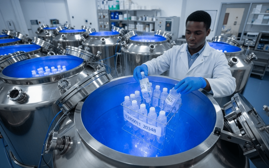
Cryopreservation of tumor tissue is the process of preserving tumor samples at extremely low temperatures, typically -80°C (dry ice freezer) or -196°C (liquid nitrogen). This halts cellular metabolism and degradation, maintaining the structural integrity and biological characteristics of the tissue for extended periods.
Why is it done? – Cryopreservation of a Tumor
Cryopreservation of tumor tissue is crucial in oncology for several reasons:
Research and Diagnostics:
- Biobanking: It allows for the creation of tumor tissue banks, providing a valuable resource for future research, including studying tumor progression, identifying biomarkers, and developing new therapies.
- Genetic Analysis: Preserved tissue retains its DNA, RNA, and other genetic information, enabling in-depth analysis of mutations and gene expression patterns that can guide personalized cancer treatments.
- Drug Testing: Cryopreserved tumor slices or cells can be used for in vitro or in vivo drug testing, helping to assess drug efficacy and develop new anti-cancer agents.
- Heterogeneity Studies: It allows researchers to study the heterogeneity within a tumor, which is important for understanding treatment resistance.
- Personalized Medicine:
- Anti-cancer Vaccines: Cryopreserved tumor samples can be used to prepare personalized anti-cancer vaccines, leveraging the patient’s own tumor components to stimulate an immune response.
- T-cell based Immunotherapy: It can facilitate the design of T-cell based immunotherapeutic treatments.
- Fertility Preservation: While not directly cryopreservation of tumor tissue, a related application in oncology is the cryopreservation of reproductive tissues (e.g., ovarian tissue, sperm, embryos) for cancer patients who face a risk of infertility due to cancer treatments like chemotherapy or radiation.
How is it done? – Cryopreservation of a Tumor
The general process of cryopreservation involves several key steps:
- Sample Collection and Preparation: Tumor tissue, often obtained through biopsy or surgical removal, is collected as quickly as possible to minimize degradation. It may be minced into smaller pieces.
- Addition of Cryoprotective Agents (CPAs): These are substances (e.g., Dimethyl sulfoxide (DMSO), trehalose) added to the tissue before freezing to reduce the formation of ice crystals, which can cause significant cell damage. CPAs work by lowering the freezing point and increasing the viscosity of the solution.
- Controlled Cooling: The tissue is cooled slowly and in a controlled manner (e.g., at a rate of -1°C per minute) to allow water to leave the cells, preventing the formation of large, damaging intracellular ice crystals. This can be achieved using specialized freezing containers or programmable freezers.
- Storage: Once cooled to very low temperatures (typically -80°C for short-term or -196°C in liquid nitrogen for long-term), the samples are stored in specialized cryopreservation tubes in ultra-cold freezers or liquid nitrogen tanks.
- Thawing and Rewarming: When needed, the cryopreserved tissue is rapidly thawed, often in a water bath, and CPAs are removed to minimize their toxicity.
Cryoablation vs. Cryopreservation of a Tumor:
It’s important to distinguish cryopreservation from cryoablation (also known as cryosurgery). While both involve extreme cold, cryoablation is a minimally invasive treatment that uses extreme cold to directly freeze and destroy cancerous cells within the body. Cryopreservation of a tumor, on the other hand, is a method of preserving tissue samples for later use, not a direct treatment for the tumor itself.

Recent Comments